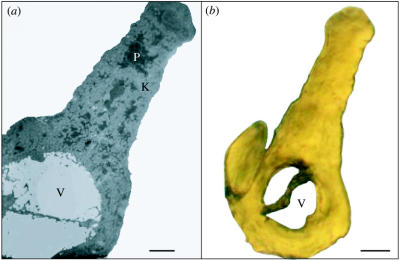
Figure 3.
(a) TEM micrograph (1900×) of a yellow American goldfinch (Carduelis tristis) feather barb. P, carotenoid pigments; K, keratin substrate; and V, air-filled vacuole. Scale bar, 1 μm. (b) Light micrograph (1000×) of a yellow American goldfinch (C. tristis) feather barb. V, Vacuole. Scale bar, 2 μm.

