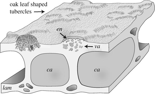
Figure 2.

Three dimensional reconstruction of the histology of Sacabambaspis based upon the material illustrated in figure 1(a–f). Reconstructed tubercle on the left includes dentine tubules based upon the interpretation of the heavily vascularized area immediately beneath the enameloid caps and comparison with Astraspis and Tesseraspis. Abbreviations: ca, cancellous middle layer; en, enameloid; lam, laminated basal layer; va, vascularized region underlying tubercles.
