
Figure 3.
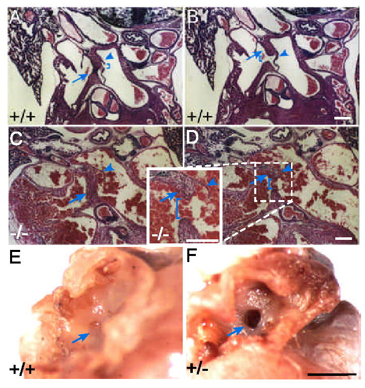
Haploinsufficiency in Ccn1 results in ASD. (A,B) WT neonatal hearts were sectioned and H & E stained. The septum primum (arrowheads) and septum secundum (arrows) were properly formed, separating the two atrial chambers. The ostium secundum (bracket in A) and fossa ovalis (bracket in B) are seen. (C,D) In Ccn1+/− pups, fusion of the septum primum (arrowheads) with the cushion tissue was unable to occur, leaving a detached septum primum and foramen ovale (brackets in D). Formation of the septum secundum (arrows) appeared normal. (E,F) In hearts of 10 month old mice, the fossa ovalis (arrow, E) was closed in the WT but the embryonic foramen ovale remained patent (arrow, F) in the Ccn1+/− mouse. White bars: 100 μm, black bar: 1 mm.