
Figure 5.
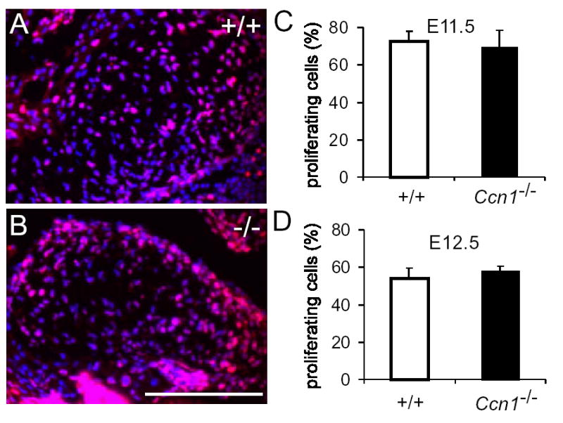
Cell proliferation in the AV cushion tissue. E11.5 (shown in A, B) and E12.5 AV cushion tissues of wild-type (+/+) and Ccn1−/− embryos were immunostained for Ki67 (red) and countered stained DAPI (blue). Results were quantified, showing equivalent proliferation rates in wild-type and Ccn1−/− embryos in E11.5 (C, n=6 each; P>0.59) and E12.5 embryos (D, n=5 each, P>0.34).