
Figure 6.
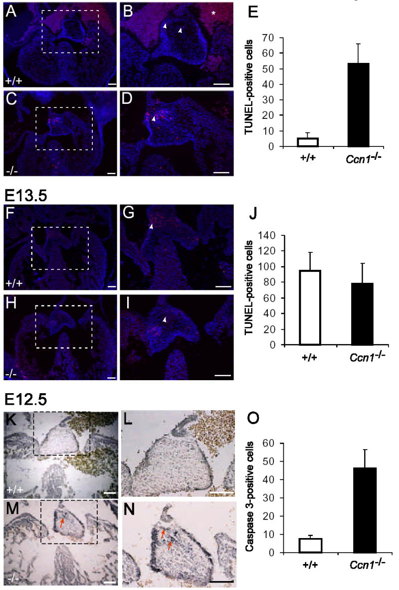
Apoptosis in the AV cushion. Apoptotic cells were identified in sections of embryonic hearts using TUNEL assay and counterstained with DAPI (A-J, arrowheads), or by immunohistochemical staining for activated caspase-3 (K-O, arrows). Higher magnifications of dashed boxes in A, C, F, H, K, and M are shown in B, D, G, I, L, and N, respectively. Precocious apoptosis was observed in E12.5 Ccn1−/− AV cushion (C,D), compared to the Ccn1+/+ cushion (A,B) by either TUNEL assay (E; P<0.001) or immunohistochemical staining of activated caspase-3 (K-O; P<0.001). At E13.5, similar numbers of TUNEL-positive cells were observed in AV cushions and valvular leaflets of both WT (F, G) and Ccn1−/− (H, I) hearts (J; P>0.30). Apoptotic cells were counted from transverse sections of AV cushions of Ccn1+/+ and Ccn1−/− hearts (each group n=5), and the average numbers of TUNEL-positive cells in 10 μm sections are shown in E, J, and O. Error bars are standard deviations. The star in B indicates background staining from red blood cells. Bars = 100 μm.