
FIGURE 4. CD80 cytoplasmic domain deletion resulted in decreased and differently localized CD80 recruitment to the IS with naïve T cells.
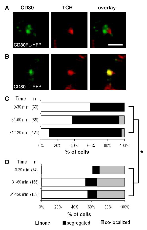
In the presence of 100 nM of MCC peptide, naïve 5C.C7 T cells and CHO cell interactions resulted in CD80FL-YFP and CD80TL-YFP molecules differentially accumulated in the IS. (A) CD80FL-YFP accumulation was segregated from the TCR at the interface of the IS while (B) CD80TL-YFP accumulation co-localized with the TCR in a central cluster in the IS. The color overlay images show TCR in red and CD80 in green. Areas of TCR and CD80 overlap are yellow. Images are cross-section of a 3-D plane rotated in an en face view with T cell on top of the CHO-APC. Scale bar = 2 μm. Quantitation of the accumulation patterns of CD80FL-YFP (C) versus CD80TL-YFP (D) in the IS over a 2 hour time course. No CD80 accumulation in the cSMAC (white), CD80 segregated from the TCR, segregation (black), and CD80 co-localized with the TCR in a central cluster, co-localized (grey). Only cells with an LFA-1 ring (pSMAC) were scored. The percentage of cells in each accumulation pattern represents the mean percentage of 3 independent experiments. The hypothesis that there was no association between cell type and pattern of accumulation was rejected with P-value less than 0.0001 (*) from the three experiments combined or each experiment individually and they each suggested significant association between cell types and patterns. The conditional independence between the “segregated” and “co-localized” and two cell types was also tested and rejected with P-value less than 0.0001 (whether or not the P-value was corrected for multiple testing).