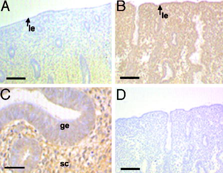
Fig. 3.

Expression of ErbB4 in human endometrium during the menstrual cycle. Tissue sections derived from proliferative (A) and secretory (B and C) endometrium were stained with anti-ErbB4 antibodies. Lumenal edge (arrow, le), glandular epithelium (ge), and stromal cells (sc) are indicated. D, Control staining was performed with antibodies preincubated with the appropriate control peptide. Scale bars, 50 μm (A, B and D) and 125 μm (C).
