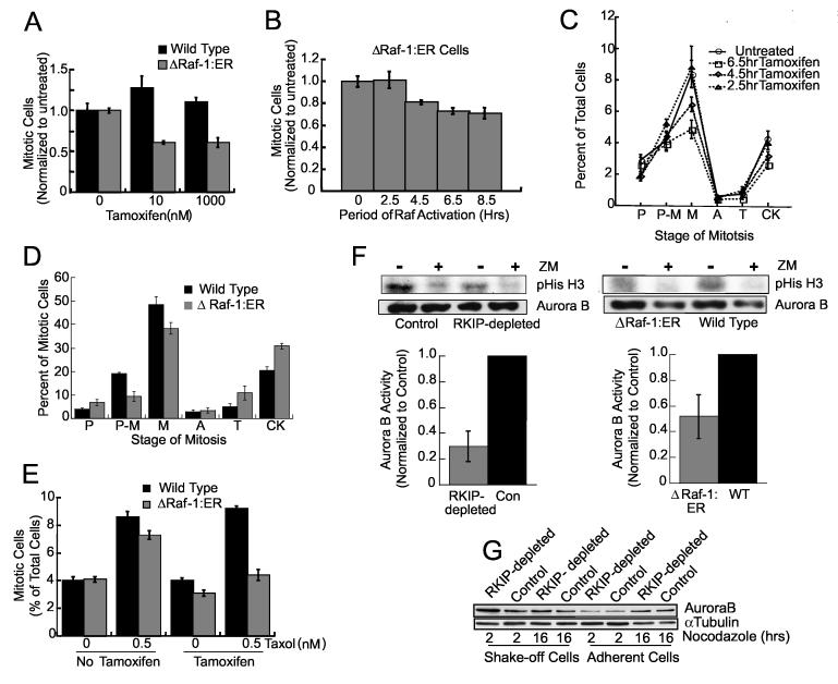
Figure 6.
Raf-1 Activation Mimics RKIP-Depletion (A) Wild type or HeLa cells stably expressing ΔRaf-1:ER were treated for 24 hrs with tamoxifen and mitotic cells counted. (B) 1 μM tamoxifen was added to synchronized ΔRaf-1:ER HeLa cells at .5, 2.5, 4.5, 6.5, or 9 hrs after release. At 9 hrs mitotic cells were counted. (C) Mitotic stages of ΔRaf-1:ER HeLa cells treated as in (B). P = prophase, P-M = prometaphase, M = metaphase, A = anaphase, T = telophase, and CK = cytokinesis. P<.03 by Student t-Test for 6.5 hr tamoxifen treatment. (D) Synchronized wild type or ΔRaf-1:ER HeLa cells were incubated for 2.5 hrs, 1 μM tamoxifen added for 6.5 hrs and mitotic cells counted. (E) Wild type or ΔRaf-1:ER HeLa cells were treated with 0 or 1 μM tamoxifen for 19 hrs, 0 or 0.5 nM Taxol added for 5 hrs and mitotic cells counted. Error bars are +/-SE. (F) RKIP-depleted and control (left), or tamoxifen-treated ΔRaf-1:ER and wild type (right) HeLa cells were analyzed for Aurora B kinase activity. ZM: 1 μM Aurora kinase inhibitor. Error bars are +/-SD (left) or range (right). (G) Synchronized RKIP-depleted or control HeLa cells were released into 200 ng/ml nocodazole. Arrested (shake-off) or adherent cells were immunoblotted for Aurora B.

