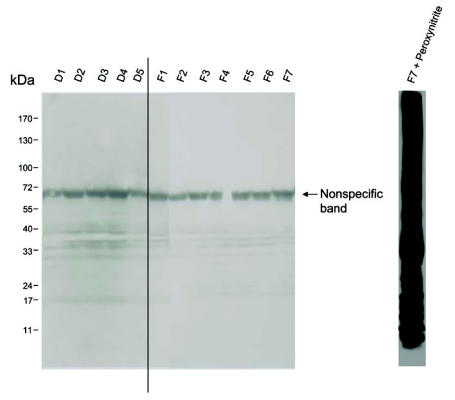
Figure 2.
Detection of protein nitration. Human ventricular tissue homogenates (F, failing; D, donor) were loaded (30 μg of protein in each lane) onto 5% to 20% gradient SDS-polyacrylamide gels and probed with a nitrotyrosine-specific antibody (1:10,000; Calbiochem). It should be noted that the major band (indicated) is recognized by the secondary antibody alone and is therefore not specific to nitrotyrosine. A positive control for nitrotyrosine staining (sample F7 treated with 500 μM peroxynitrite for 5 min) is shown on the right.

