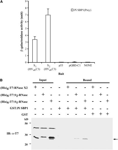
Figure 4.
Analyses of Interactions of Pi SBP1 with Full-Size and Truncated S1- and S2-RNases.
(A) Yeast two-hybrid assay. The assay was performed as described in the legend to Figure 2A. S1(HVabC3) and S2(HVabC3) are truncated S1- and S2-RNase, respectively, each containing the two hypervariable regions and the conserved C3 region. All of the negative controls are as described in the legend to Figure 2A.
(B) In vitro binding assay. The interactions between GST:Pi SBP1 and purified (His)6:T7-tagged S1-RNase, S2-RNase, and RNase X2 were analyzed as described in the legend to Figure 2B. The bound proteins (arrow) were detected by immunoblotting (IB) using an anti-T7 antibody. The band above the (His)6:T7-tagged protein in each input lane is an E. coli protein that copurified with the tagged protein.

