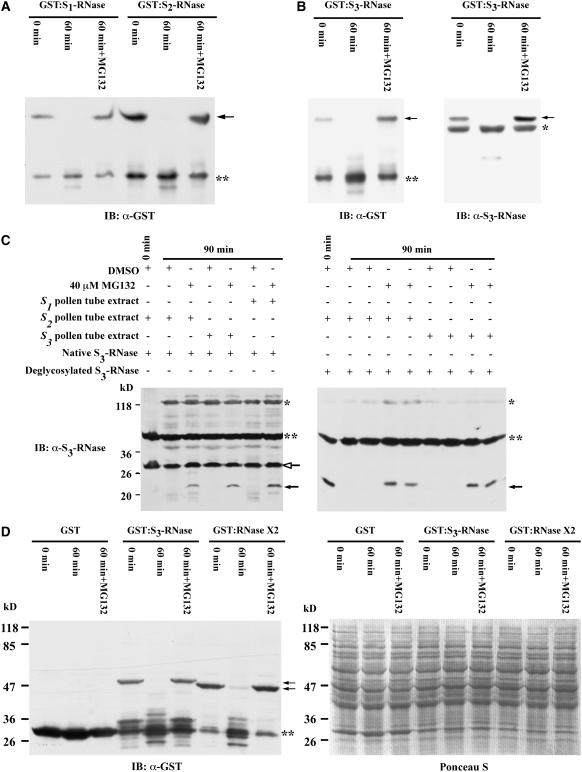
Figure 7.
Degradation Assay of Bacterially Expressed GST, GST:S1-RNase, GST:S2-RNase, GST:S3-RNase, and GST:RNase X2, as Well as S3-RNase Purified from Pistils, by Extracts of S1, S2, or S3 Pollen Tubes.
(A) GST:S1-RNase and GST:S2-RNase. Purified GST fusion proteins (0.3 μg each) were incubated separately with extracts of in vitro–germinated S2 pollen tubes in either the absence or presence of 40 μM MG132 (a specific inhibitor of the 26S proteasome) for 1 h at 30°C. An anti-GST antibody was used to detect GST:S1-RNase and GST:S2-RNase (arrow) as well as GST (double asterisks). IB, immunoblot.
(B) GST:S3-RNase. Purified GST:S3-RNase (0.3 μg) was used in the assay as described for (A), except that both anti-GST and anti-S3-RNase antibodies were used to detect GST:S3-RNase. The arrow indicates GST:S3-RNase, the single asterisk indicates a cross-reacting protein present in the reaction mixture, and the double asterisks indicate GST.
(C) Native glycosylated S3-RNase and its deglycosylated form. S3-RNase (0.1 μg) purified from pistils of the S3S3 genotype (left panel) and an equal amount of purified deglycosylated S3-RNase (right panel) were incubated separately with extracts of S1, S2, and S3 pollen tubes in either the absence or presence of MG132 (40 μM) for 90 min at 30°C. The anti-S3-RNase antibody was used to detect both glycosylated (open-headed arrow) and deglycosylated (closed arrows) S3-RNase. Single and double asterisks indicate cross-reacting proteins in the reaction mixture. A longer exposure time was used for the blot shown in the left panel (5 min) than that shown in the right panel (30 s).
(D) GST:S3-RNase, GST:RNase X2, and GST. Each purified protein (0.3 μg) was used in the assay as described for (A). Left panel, immunoblot. The arrows indicate GST:S3-RNase or GST:RNase X2, and the double asterisks indicate GST. Right panel, Ponceau S staining of the blot shown in the left panel before immunoblotting.

