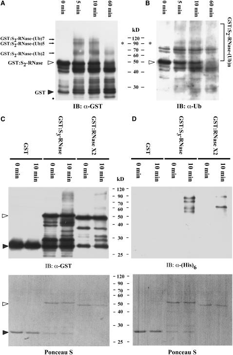
Figure 8.
Ubiquitination Assay of GST, GST:S2-RNase, GST:S3-RNase, and GST:RNase X2 by Extracts of S2 Pollen Tubes.
(A) and (B) Time course of ubiquitination and degradation of GST:S2-RNase. GST:S2-RNase (0.5 μg) was used in each reaction containing ubiquitin, and the reaction was stopped at different time points as indicated. The anti-GST antibody was used in the protein gel blot shown in (A), and an anti-ubiquitin antibody was used in the blot shown in (B). The open triangles indicate GST:S2-RNase, and the closed triangle indicates GST. The black dot indicates the degradative products of GST:S2-RNase. The closed arrows indicate three ubiquitinated forms of GST:S2-RNase with two, five, and seven ubiquitin subunits, and the asterisks indicate GST:S2-RNase conjugated with five ubiquitin subunits that was detected by both anti-GST and anti-ubiquitin antibodies. IB, immunoblot.
(C) and (D) Ubiquitination assay of GST, GST:S3-RNase, and GST:RNase X2. Purified GST, GST:S3-RNase, and GST:RNase X2 (0.5 μg each) were used in the assay as described for (A) and (B), except that the reaction contained (His)6:ubiquitin and was analyzed only at the 10-min time point. Each reaction mixture was then divided equally and electrophoresed on two duplicate gels. The transferred membranes were immunoblotted with the anti-GST antibody (C) and the anti-(His)6 antibody (D). Top panels, immunoblot. Bottom panels, Ponceau S staining of the blots shown in the top panels before immunoblotting.

