Figure 3.
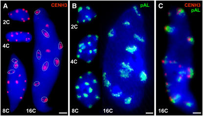
Number and Appearance of EYFP-CENH3–Specific Immunosignals and Centromere-Specific FISH Signals in A. thaliana Nuclei of Different Endopolyploidy Levels.
Sorted nuclei after immunofluorescence labeling of EYFP-CENH3 (A), FISH detection of centromeric ∼180-bp repeats (B), and the combination of both in a 16C nucleus (C). DNA is counterstained with DAPI (blue). In 2C to 8C nuclei, immunofluorescence and FISH signals are mainly compact, while in 16C, signals were often disperse or split into smaller foci (circled). Bars = 2 μm.

