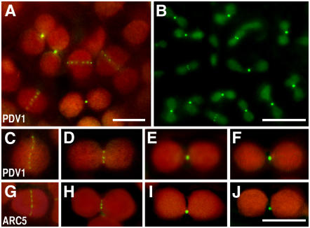
Figure 4.
Localization of GFP-PDV1 and GFP-ARC5.
A GFP-PDV1 transgene was expressed in pdv1-1 plants, and GFP fluorescence was observed by fluorescence microscopy. The fluorescence signal of GFP is green, and the autofluorescence of chlorophyll is red. The dim green fluorescence in (B) is background, because similar fluorescence was observed without the GFP-PDV1 transgene. Expression of the GFP-PDV1 transgene complemented the mutant phenotype of pdv1-1. Bars in (A), (B), and (J) = 5 μm. (C) to (J) are shown at the same magnification.
(A) Chloroplasts in mesophyll cells of a young leaf expressing GFP-PDV1.
(B) Plastids in fringes of petals from flower buds expressing GFP-PDV1.
(C) to (F) Comparison of GFP-PDV1 localization among chloroplasts at various stages of division.
(G) to (J) Comparison of GFP-ARC5 localization among chloroplasts at various stages of division.

