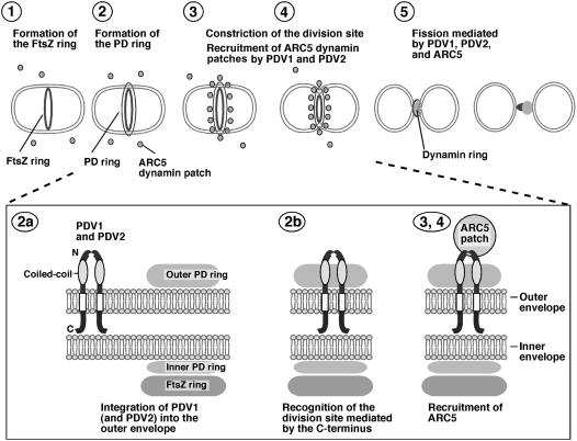
Figure 9.
Working Model of PDV1 and PDV2 Function during Plastid Division.
The top panel shows an abbreviated model of the plastid division pathway. (For additional details without PDV1 and PDV2, see Miyagishima et al., 2003b; Osteryoung and Nunnari, 2003.) The FtsZ ring, inner plastid–dividing (PD) ring, and outer plastid–dividing ring assemble sequentially (steps 1 and 2) before the onset of constriction (step 3). During constriction, the ARC5 dynamin patches are recruited from the cytosol to the division site by PDV1 and PDV2 (steps 3 and 4) to form a ring structure during a late stage of division (step 5). The bottom panel shows details of proposed PDV1 and PDV2 function. PDV1 (and probably PDV2) is integrated into the outer envelope membrane (step 2a), perhaps interacting via their coiled-coil domains. Localization of PDV1 (and probably PDV2) to the midplastid is mediated at least in part by the conserved Gly at the C terminus, which may be oriented toward the intermembrane space (but see Discussion) (step 2b). PDV1 (and probably PDV2) forms a discontinuous ring structure in the outer envelope membrane; this ring is required for the recruitment of ARC5 dynamin patches to the cytosolic surface of the outer envelope membrane (step 3).

