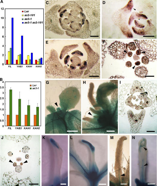
Figure 5.
Expression of the Leaf Polarity Marker Genes.
(A) and (B) Analyses by real-time RT-PCR for transcript levels of FIL, YAB3, KAN1, and KAN2 in wild-type Ler, as2-101, ae3-1, and ae3-1 as2-101 rosette leaves (A) or in the Ler and ae3-1 cauline leaves (B). Bars show standard error.
(C) to (F) In situ hybridization of FIL expression in Ler (C), as2-101 (D), ae3-1 (E), and ae3-1 as2-101 (F).
(G) and (H) Analyses of REV expression using an rev-9 enhancer trap line. Note that only wild-type and ae3-1 as2-101 mutant plants that are hemizygous for rev-9 were characterized. Shoot apices from 12-d-old wild-type Ler (G) or ae3-1 as2-101 (H) are shown.
(I) and (J) In situ hybridization of REV expression in ae3-1 (I) and ae1-3 as2-101 (J).
(K) to (N) Needle-like leaves of ae3-1 as2-101. Note that these needle-like leaves show varying degrees of adaxial/abaxial differentiation and vascular tissue formation. Arrowheads in (M) and (N) show GUS staining, indicating the insufficiently developed vascular tissues.
Bars = 100 μm in (C) to (F), (I), and (J), 200 μm in (G), (H), and (K) to (M), and 500 μm in (N).

