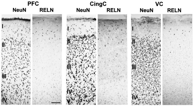
Figure 1.
Photomicrographs of 20-μm sections showing the distribution of NeuN- and Reln-expressing cells in the PFC, CingC, and VC of the baboon. Note that immunostaining for Reln strongly labels a small population of cells predominantly located in layer I. In addition to these Reln-expressing cells, there appears to be a faint immunolabeling of Reln on many pyramidal cells in deeper cortical layers. (Calibration bar = 200 μm.)

