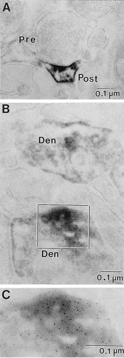
Figure 5.

Electron micrographs of ultrathin sections of Reln immunolabeling in the patas PFC layers I and II. (A) Immunoperoxidase reaction product in a dendritic spine is concentrated in the area of the postsynaptic density as well as the surrounding dendritic plasma membrane. (B) Double-immunolabeling showing the colocalization of Reln (immunoperoxidase-diaminobenzidine) and integrin α3-subunit (silver-enhanced 1-nm ImmunoGold) in ultrathin sections from patas PFC layers I and II. Both dendritic processes present in this electron micrograph are immunopositive for Reln and integrin α3-subunit. (C) Higher magnification of the area outlined in B. Pre, presynaptic bouton with synaptic vesicles; Post, postsynaptic spine; Den, dendritic process. (Magnification: A = ×75,000; B = ×125,000; C = ×212,500.)
