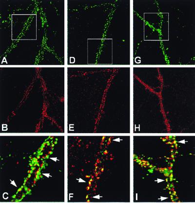
Figure 4.
Immunocytochemical localization of ErbB-4 at synaptic sites of cultured hippocampal cells. Double immunofluorescence was performed on dissociated hippocampal neurons cultured for 3 wk. (A–C) ErbB-4 receptor puncta (red) are located at synaptic sites apposed to a subset of the synaptophysin-positive presynaptic terminals (green). The yellow color in the enlarged view (C) indicates the extent of immunofluorescence overlap. (D–F) ErbB-4 receptors (red) and PSD-95 (green) colocalize in a subset of synaptic puncta (yellow). (G–I) ErbB-4 receptors (red) and the NR1 subunit of the NMDA receptor (green) also colocalize at puncta (yellow). Areas that are boxed are enlarged (Bottom), and the arrows indicate examples of colocalization.

