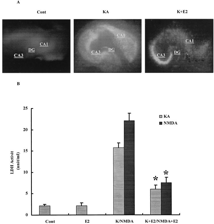
Figure 1.
Effects of E2 on KA- and NMDA-elicited neurotoxicity. Hippocampal slices were prepared as described in Materials and Methods and were maintained in cultures for 10–14 days. They were treated with NMDA or KA (50 μM) for 3 h and returned to normal medium for 24 h. When present, E2 was added at 1 nM 24 h before NMDA or KA and was present for the duration of the experiment. (A) PI uptake in control, 24 h after a 3-h treatment with KA (50 μM) in the absence or presence of E2. DG, dentate gyrus. (B) LDH activity in medium measured in control (Cont), 48 h after treatment with E2 (E2), 24 h after KA or NMDA treatment in the absence (K/NMDA) or presence of E2 (K+E2/NMDA+E2). Results are means ± SEM of six to eight experiments. *, P < 0.001 compared with KA or NMDA (Student's t test).

