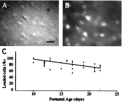
Figure 1.
Loading mature cortical slices with fura-2 AM. DIC (A) and fluorescence (B) images of the lower layers of a visual cortex slice from a P10 mouse with the pial surface to the bottom right. The slice was loaded with fura-2 AM. Somata and apical dendrites from many neurons are fluorescent, and pyramidal and nonpyramidal neurons label equally. (Scale bar = 20 μm.) (C) Percentage of loaded neurons as a function of the age of the animal. Each point represents one brain slice. Loading is close to 100% at P10, but it declines to ≈70% of the neurons at P20. The line represents the best linear fit.

