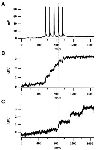
Figure 2.
Follower neuron time-locked to the stimulation of a trigger cell. (A) Whole-cell recording of neuron 1 (trigger). In response to five depolarizing current pulses (3 nA; 5 msec), the neuron fires five APs. (B) Simultaneous fluorescence measurements with a photodiode of the somatic region of neuron 1 show discrete calcium accumulations that correspond to the five APs. The sign of the fluorescence signals in all figures has been inverted. ADU, analog-digital voltage units. (C) Simultaneous fluorescence measurements of neuron 2 (follower), showing an intracellular calcium concentration ([Ca2+]i) transient phase-locked with the fourth AP of neuron 1. The onset of the calcium signal in neuron 2 occurs coincident (<0.6 msec) with the peak of the AP in neuron 1. Neuron 2 has subsequent [Ca2+]i increases. The experiment was carried out in a P18 mouse cortical slice under ACSF with 2 mM Ca2+/1 mM Mg2+.

