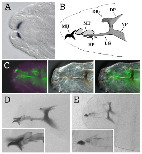
Figure 1.
kal-1 overexpression in the cells responsible for the formation of the cephalopharyngeal skeleton causes alterations of the sclerotized components of the head. (A) Stage 16 wild type embryonic head. The kal-1 transcript is detected in the region of the gnathal lobes that will give rise to the anterior part of the cephalopharyngeal skeleton. (B) Schematic representation of the cephalopharyngeal skeleton of a L1 Drosophila larva. MH mouth hook, MT median tooth, HP H-piece, LG lateralgräten, DBr dorsal bridge, DP dorsal process, VP ventral process. (C) Fluorescence (left), bright-field (center), and merged (right) images of the expression in the cephalopharyngeal skeleton of a stage 17 embryo of the UAS-GFP reporter, driven by the 179y-Gal4 line. (D) Wild type stage 17 embryonic head cuticle. In the inset, a magnification of the mouth hooks. (E) Head cuticle of a stage 17 179y-Gal4/+; UAS-kal-1/+; UAS-kal-1/+ embryo. The head skeleton structure appears less sclerotized; principally the median tooth, the H-piece, and the lateralgräten, but also the dorsal bridge, the dorsal process, and the ventral process appear thinner than in the wild type. The mouth hooks lack the posterior part (inset).

