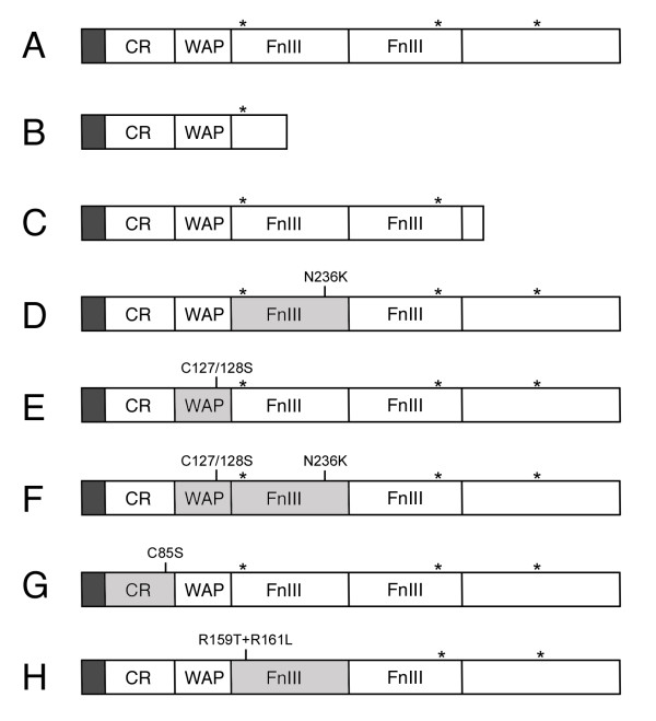
Figure 2.
Schematic structure of Dmkal-1 and of the different mutant proteins analyzed. (A) Wild type Dmkal-1. (B) Dmkal-1 [W200-STOP]. (C) Dmkal-1 [H384-STOP]. (D) Dmkal-1 [N236K]. (E) Dmkal-1 [C127/128S]. (F) Dmkal-1 [C127/128+N236K]. (G) Dmkal-1 [C85S]. (H) Dmkal-1 [R159T+R161L]. CR, Cysteine-rich domain; WAP, Whey Acidic Protein-like domain; FnIII, Fibronectin-like type III domain. Asterisks indicate the position of heparan-sulfate binding site consensus sequences. In gray the domains affected by mutations.

