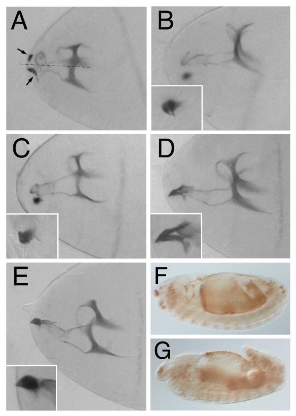
Figure 3.
Cephalopharyngeal skeleton phenotypes induced by overexpression of the different DmKal-1 mutant proteins. (A, B, C, D, E) Head cuticle of stage 17 embryos. (A) Stage 17 179y-Gal4/+; UAS-kal-1 [N236K] embryo. The head skeleton structure appears less sclerotized and the median tooth is altered. Moreover, the mouth hooks (arrows) lack the posterior part and appear distant from the mid-line (dotted line). (B) Stage 17 179y-Gal4/+; UAS-kal-1 [C127/128S] embryo. (C) Stage 17 179y-Gal4/+; UAS-kal-1 [C127/128S+N236K] embryo. (D) Stage 17 179y-Gal4/+; UAS-kal-1 [C85S] embryo. The mouth hooks base shows a reduction of the dorsal process. (E) Stage 17 179y-Gal4/+; UAS-kal-1 [R159T+R161L] embryo. In the insets, a magnification of the hooks. (F, G)) Stage 13 embryos immunostained with anti-Hind antibody. (F) 179y-Gal4/+; UAS-kal-1; UAS-kal-1 embryo. GBR occurs normally. (G) 179y-Gal4/+; UAS-kal-1 [N236K] embryo. The embryo shows an incomplete GBR process.

