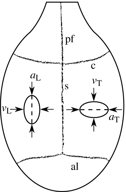
Figure 1.
Schematic of the rat cranial vault, showing the approximate location of the longitudinal and transverse incisions. This dorsal view shows the posterior frontal (pf), sagittal (s), coronal (c) and anterior lambdoidal (al) sutures. The anterior–posterior and medial–lateral dashed lines indicate the approximate locations at which the respective longitudinal and transverse incisions were made in the dura mater. The anatomical side of the incisions was randomized for each rat. The solid ovals illustrate how the edges of the incision often retracted, causing the incision to assume an elliptical shape. The length (a) and maximum width (v) of each incision was measured as shown.

