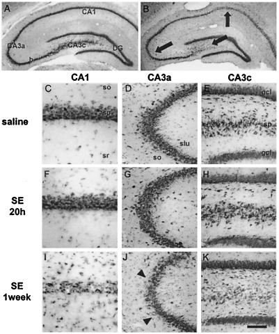
Figure 1.
Status epilepticus (SE) induces delayed neurodegeneration of CA1 and CA3 pyramidal cells. Histological analysis of toluidine blue-stained brain sections at the level of the dorsal hippocampus. Control brain from an animal sacrificed 7 days after saline injection (A and C–E). There is no detectable neuronal damage 20 h after KA administration (F–H), but at 1 week, loss of CA1 and CA3 pyramidal neurons is prominent (B and I–K). Arrows in B indicate location of higher magnification views in C–K. Arrowheads in J indicate a region of extensive necrosis. Many neurons in I show pyknotic nuclei. (Scale bar: 3,690 μm in A and B; 150 μm in D, E, G, H, J, and K; 75 μm in C, F, and I.)

