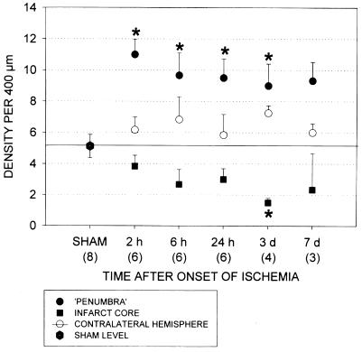
Figure 8.
The density of STC-stained neuronal processes was histologically quantitated in the cerebral cortex in three locations of the infarcted rat brains: the ischemic core, the narrow rim surrounding the infarct (“penumbra”), and the homologous area in the contralateral, nonischemic hemisphere. Corresponding cortical areas were examined in sham-operated animals. An investigator blinded to the source of the samples counted the numbers of vertically oriented STC-stained neuronal processes cut by a 400-μM bar placed horizontally on the cerebral cortex. The data are expressed as mean ± SE. Statistical tests were performed according to one-way ANOVA followed by Dunn's post hoc test. *, P < 0.05 for the given number of animals in each group.

