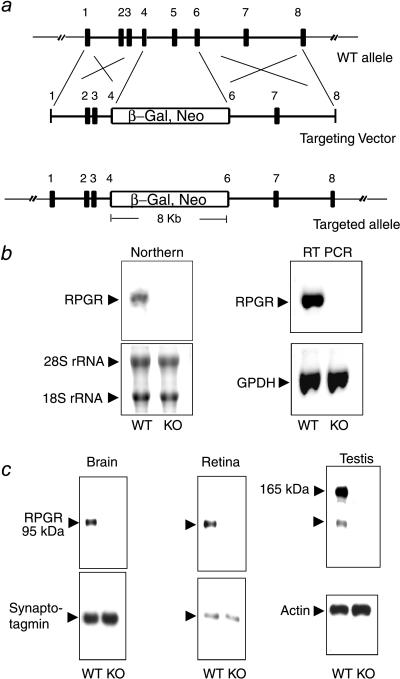
Figure 1.
Targeted disruption of the RPGR gene. (a) The targeting construct. Partial structure of the WT allele is shown (exons 1–8; filled boxes). The targeting vector contains a selectable marker (β-Gal, Neo) that replaces part of exon 4 through part of exon 6. (b) Absence of the normal RPGR transcript in the KO mice. (Left) Northern blot analysis with a full-length RPGR cDNA probe could detect a 3-kb transcript in brain RNA from WT but not from KO mice. Fluorescent dye-stained rRNA bands are shown as a loading control. (Right) Reverse transcription–PCR analysis confirms the absence of the normal RPGR transcript in the KO mice. PCR amplification of GPDH from the same cDNA templates is shown as a control. (c) RPGR protein expression was ablated in the KO mice. Total proteins from brain, retina, or testis were probed with anti-RPGR antibodies on immunoblots. The same blots were reprobed with either synaptotagmin (abundant in neural tissues) or actin antibodies as loading controls.

