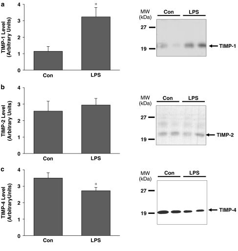
Figure 6.
(a) TIMP-1, (b) TIMP-2 and (c) TIMP-4 protein content in aortae excised from lipopolyssaccharide (LPS) or vehicle (Con) – treated rats. Right panels show representative immunoblots taken from aortae from two control rats (Con) and two LPS-treated rats (LPS). TIMP-3 was not detectable (data not shown). Position of molecular weight markers is depicted on the left (*P<0.05, independent samples t-test, n=12–13 rats/group for TIMP-1 and TIMP-4, n=6 rats/group for TIMP-2).

