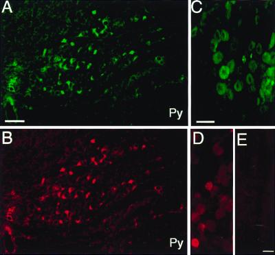
Figure 1.
Immunohistochemistry controls. Immunostaining is shown in the inferior olive by using an antibody to VR1 receptor raised in guinea pig (A) and rabbit (B) in adjacent sections. Note that the cells immunopositive with both antibodies are identical. (C and D) Immunostaining using the same antibodies (C, guinea pig; D, rabbit) in the DRG. (E) A section of DRG immunostained with the rabbit antibody that had been previously preabsorbed with the immunizing peptide (negative control). Py, pyramidal tract. (Scale: 100 μm, A and B; 50 μm, C–E.)

