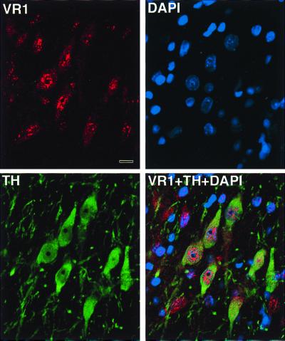
Figure 2.
Fluorescent images taken through a confocal microscope. Section thickness: 1 μm. Rat substantia nigra immunostained with the anti-VR1 antibody raised in rabbit and then anti-rabbit IgG-Alexa 546 dye was applied as a secondary antibody. Many of the substantia nigra neurons show a punctate-like cytoplasmic as well as nuclear staining. A monoclonal mouse anti-TH antibody (Boehringer Mannheim) was used to mark the dopaminergic neurons in the same area. FITC was used as a secondary antibody-green fluorescence and 4′,6-diamidino-2-phenylindole (DAPI) (blue) was used as a nuclear marker viewed through the UV filter. Finally, a digital overlay of the three images demonstrates that all substantia nigra TH immunopositive neurons are also VR1 positive and shows that some of the immunostaining is nuclear. Note that there are some VR1 immunopositive cells that are not TH positive. (Scale: 10 μm.)

