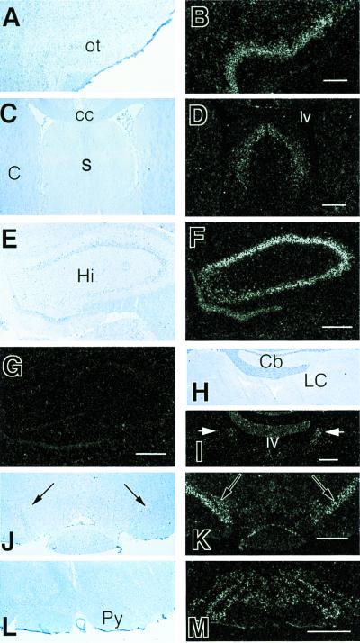
Figure 4.
ISHH in the rat CNS using a riboprobe complementary to the VR1 mRNA. We found high to medium levels of mRNA being expressed in neurons of several areas including the olfactory cortex (A) bright-field and (B) dark-field illumination of the same field; the lateral and dorsal septal nuclei (C) bright-field and (D) dark-field illumination; hippocampal pyramidal cells and dentate gyrus (E) bright field and (F) dark field; in the locus coeruleus (arrows) (H) bright field and (I) dark field; in the substantia nigra (J) bright field and (K) dark field, and in the inferior olive (L) bright-field and (M) dark-field illumination. (G) A hippocampal area similar to E and F that was hybridized with a sense probe and shows no signal. ot, olfactory tubercle; cc, corpus callosum; s, septum; lv, lateral ventricle; C, caudate; Cb, cerebellum; Hi, hippocampus; LC, locus coeruleus; Py, pyramidal tract. (Scale: 1 mm, A–D, L, and M; 800 μm, E–G, J, and K; 500 μm, H and I).

