
Figure 8.
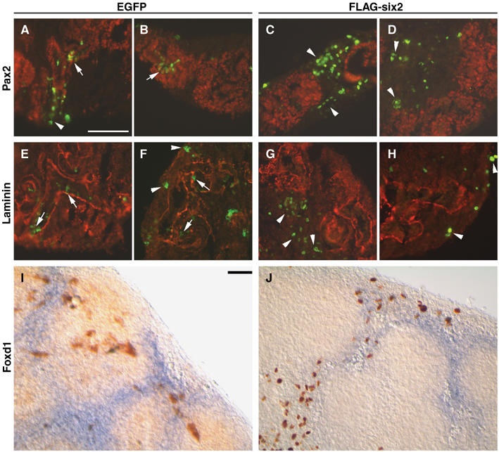
Overexpression of Six2 in wild-type kidney organ cultures. Forty-eight hours after microinjection and electroporation of EGFP or FLAG-Six2 expression plasmids, sections of E12.5 kidney organ cultures were labeled with antibodies specific for Pax2 (red; A–D), laminin (red; E–H), EGFP (green), or FLAG-Six2 (green). (A, B) EGFP and Pax2 were coexpressed in epithelial structures (arrows), and EGFP was also expressed in peripheral mesenchyme (arrowhead). (C, D) Cells expressing FLAG-Six2 were almost exclusively found in peripheral and interstitial mesenchyme (arrowheads), separated from Pax2-positive cells. (E, F) EGFP-positive cells were located within the developing tubules (arrows), as demarcated by laminin-containing basement membranes, and in the peripheral mesenchyme (arrowheads). (G, H) FLAG-Six2-expressing cells (arrowheads) were not surrounded by laminin-containing basement membranes and exhibited a mesenchymal phenotype. (I, J) In situ hybridization for Foxd1 followed by immunohistochemistry using anti-GFP (I) or anti-FLAG (J) antibodies indicated that the cells expressing FLAG-Six2 resided mainly in the interstitial stroma, whereas cells expressing the EGFP control vector resided in all cell populations. Scale bar, 100 μm.