
Figure 4.
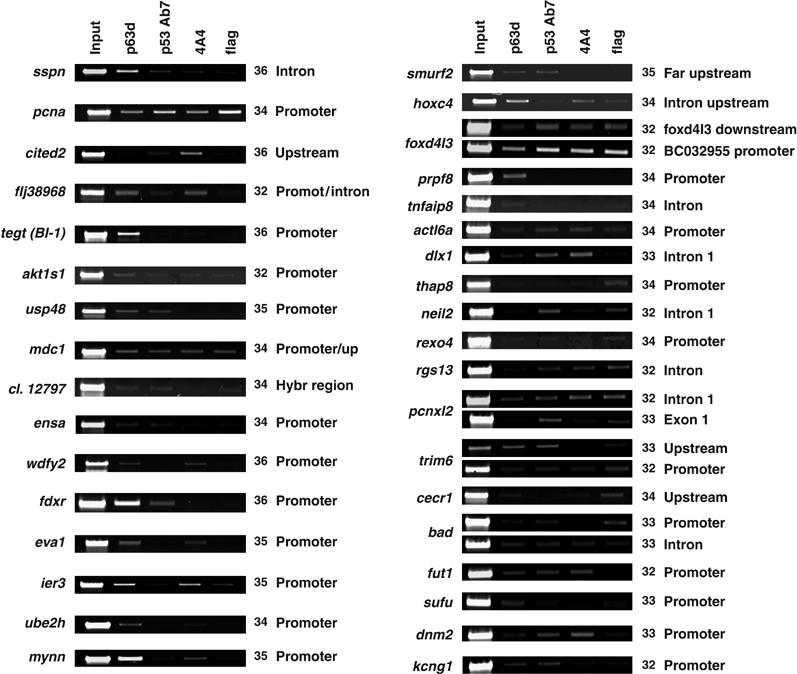
Validation of targets in human primary KCs. ChIP from human primary KCs performed with α-p63 (lane 2), α-p53 (lane 3), α-p63 monoclonal 4A4 (lane 4) and control α-Flag (lane 5) antibodies. The same layout of Figure 2 was maintained, with the strong positive loci observed in HaCaT cells on the left side, and the weak ones on the right. The position of the primers and PCR cycles are indicated.