
Figure 8.
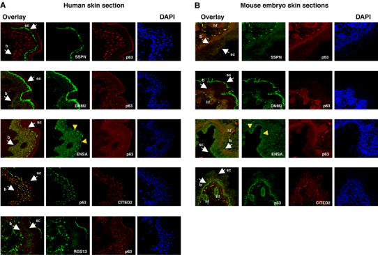
Immunostaining of human and mouse skin sections. Confocal images of adult human skin (A) and mouse 19.5-day-old embryo skin sections (B), immunostained with the indicated primary antibodies. For each target, costaining with p63, nuclear staining with DAPI and the merge (overlay) of the two images are indicated. b: basal layer; sc: subcorneum layer; hf: hair follicles (mouse only). Yellow arrowheads in the ENSA panels indicate the perinuclear accumulation of the protein in the external layers.