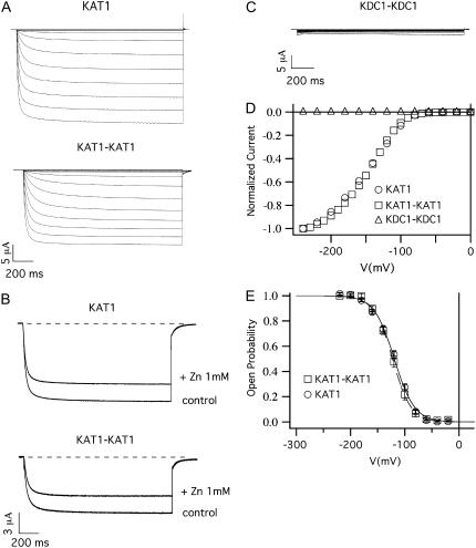
FIGURE 3.
KAT1-KAT1 dimer (but not KDC1-KDC1 dimer) forms functional channels. (A) Functional expression of the KAT1-KAT1 dimeric construct. Ionic currents elicited by voltage steps from 0 mV to −240 mV in −20 mV decrements from a holding potential of 0 mV in oocytes injected with KAT1 and KAT1-KAT1 tandem constructs. (B) Inhibition of the current for the KAT1 and KAT1-KAT1 subunits, on the addition of 1 mM of ZnCl2 to the bath solution. Holding, step, and tail potential are, respectively, 0 mV, −160 mV, and −50 mV. (C) Xenopus oocytes injected with KDC1-KDC1 tandem and stimulated with the same potential protocol used in panel A did not display any current. (D) Current voltage characteristics of the current families in panels A and C were normalized to the steady-state currents at −240 mV and plotted as a function of the applied voltage. (E) Open-probability characteristics of the KAT1 monomeric and KAT1-KAT1 dimeric channels. Data were obtained from steady-state currents like those shown in the upper panel plotted versus voltage. Continuous and dashed curves are the best fit of mean open probability (mean ± SE) obtained from at least eight experiments on KAT1 and KAT1-KAT1 channels, respectively.

