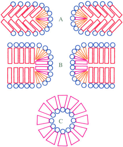
FIGURE 8.
Schematics of the energetically favorable localization of different lipids in short and long pores. (A and B) Cross section of short and long pores orthogonal to the plane of membrane. (C) Cross section of the middle part of the pore in plane of membrane. Lamellar lipids are distinguished by red cylinders in hydrophobic region; lipids with positive intrinsic curvature are distinguished by coral cones; lipids with negative intrinsic curvature are distinguished by magenta inverted cones.

