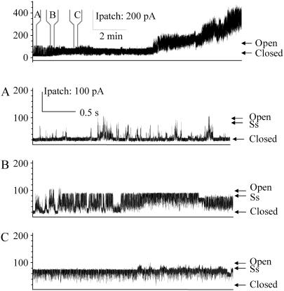
FIGURE 2.
G26C locks in an open channel conformation when modified by MTSET placed on the periplasmic side. The uppermost trace shows G26C activity with MTSET added by backfilling the pipette as described in Materials and Methods, note that no pressure was applied before or during this trace. At the time points indicated by panels A, B, and C, the trace has been expanded. Panel A shows the first channel starting to open and the preference for substates and short open dwell times. Panel B shows the first channel being locked into an open state. Panel C shows the final preference of the channel for a common 4/5ths open substate (labeled as Ss).

