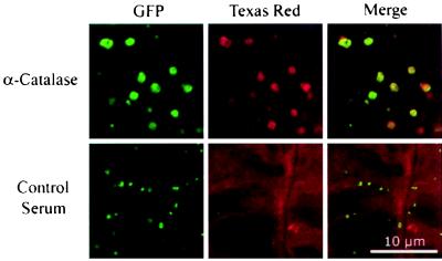
Figure 3.
Torus structures react with an antibody directed against a peroxisomal protein. Shown are confocal images of whole-mount Arabidopsis seedling tissue probed with anticatalase serum (catalase, upper row) and serum from a control rabbit (serum control, lower row). EGFP fluorescence was detected at 510–532 nm with excitation at 488 nm (GFP, shown in green pseudocolor). Texas Red-conjugated secondary antibody was detected at 595–615 nm with excitation at 568 nm (Texas Red, shown in red pseudocolor). Correspondence of the fluorescence patterns is shown in color-merged images (Merged).

