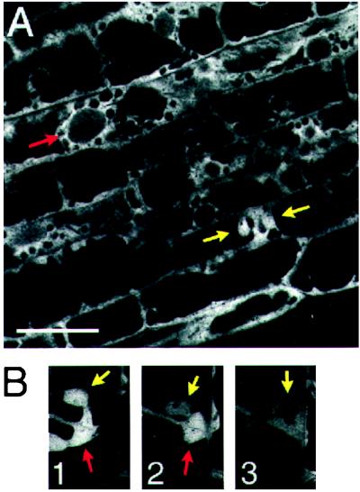
Figure 4.
GFP∷GF14 shows dynamic nuclear localization. (A) Confocal image of hypocotyl cells expressing GFP∷GF14. GFP∷GF14 localizes to the nuclei of cells undergoing cytokinesis (yellow arrows) but not to the nuclei of most interphase cells (red arrow). (B) Confocal time series of hypocotyl cell undergoing cytokinesis. GFP∷GF14 accumulates in the nuclear lumen early in cytokinesis (1, yellow arrow) and dissipates as cytokinesis proceeds (2 and 3, yellow arrows). Part of the phragmosome, the site of synthesis of the new cell wall, is visible as a mass of cytosolic fluorescence (red arrows). Note that the intensity of signal decreases in the nucleus, but fluorescence remains relatively constant in the cytoplasm at the periphery of the cell. The phragmosome, brightly labeled at the start of the sequence, dissipates by the third frame. The frames were acquired at t = 0, 17, and 22 min, respectively. (Bar = 20 microns.)

