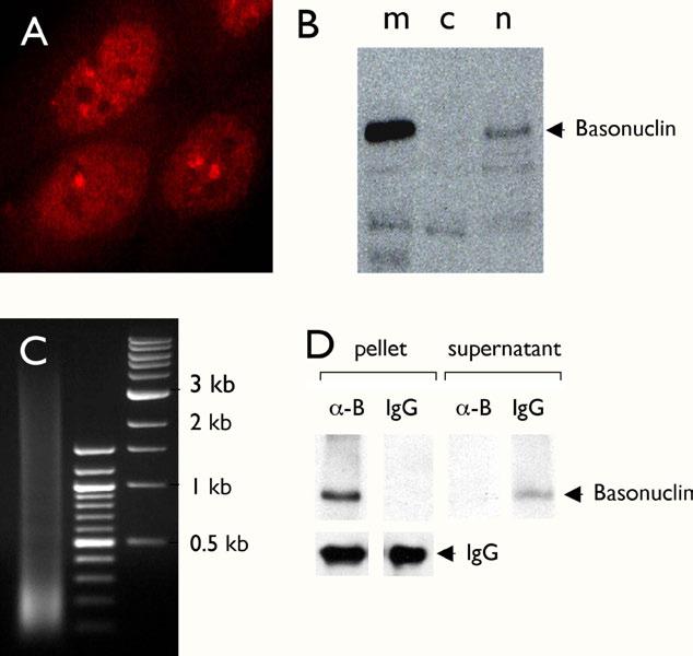
Figure 4.

ChIP assay in HaCaT cells. Immunocytochemical staining of cultured HaCaT cells with an anti-basonuclin antibody showed that basonuclin was localized mainly in the nucleoplasm with aggregates within nucleolus and the perinucleolar region (A). Basonuclin’s nuclear localization was confirmed by Western analysis of cytoplastic (B, lane c) and nuclear (B, lane n) fractions. Lane m, exogenously expressed basonuclin in the 293 cells, serving as a molecular weight marker. For the ChIP assay, the crosslinked chromatin was sonicated to produce DNA fragments of size between 0.1 to 3.5 kb (Lane 1, C). Lanes 2 and 3 in C, molecular weight markers. Basonuclin in the ChIP lysate was quantitatively precipitated (i.e., only in the pellet, not in the supernatant) with the anti-basonuclin antibody (∼-B) but not with the control rabbit IgG (D). The lower left panels show that similar amounts of antibodies were used in the assay (IgG).
