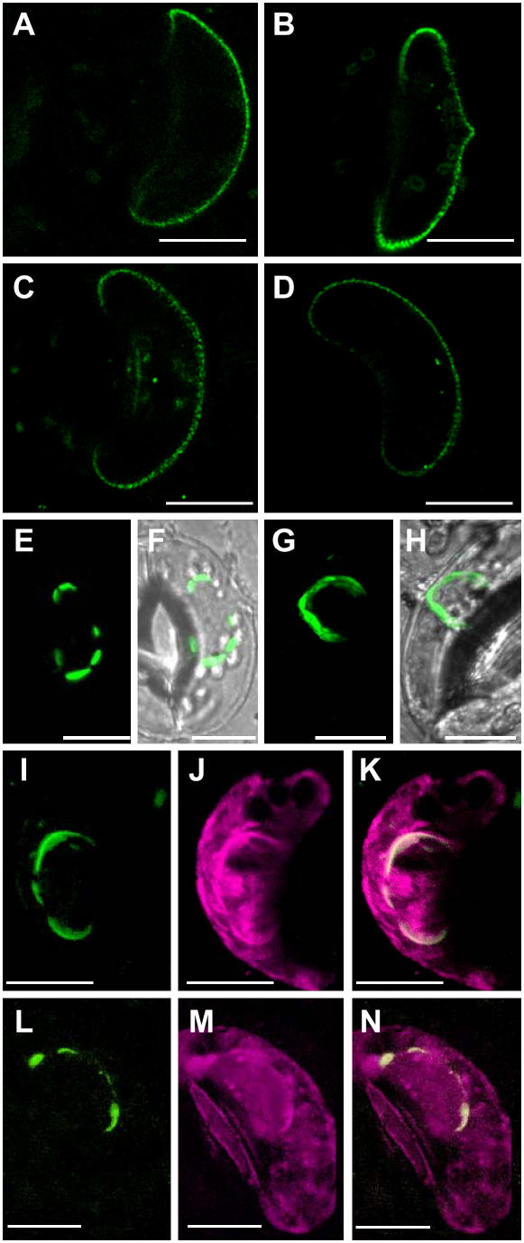
Figure 3.
Mutation of the diacidic motif (I) of KAT1 results in ER retention. A to D, Projection of two optical sections through the equatorial region of guard cells expressing KAT1∷GFP (A), KAT1(II)∷GFP (B), KAT1(III)∷GFP (C), or KAT1(IV)∷GFP (D). E to H, Overlay of transparency and fluorescent projection of four optical sections through the equatorial region of a guard cell expressing KAT1(I)∷GFP (E and F) or wild-type KAT1∷GFP and GDP-fixed Sar1 (G and H). I to K, Projection of four optical sections through the equatorial region of a guard cell cotransfected with KAT1(I)∷YFP and CFP∷HDEL; KAT1(I)∷YFP fluorescence is shown in I, CFP∷HDEL fluorescence is shown in J, and K represents overlay of both images. L to N, Projection of four optical sections through the equatorial region of a guard cell cotransfected with GDP-fixed Sar1, KAT1∷YFP, and CFP∷HDEL; KAT1∷YFP fluorescence is shown in L, CFP∷HDEL fluorescence is shown in M, and N represents overlay of both images. Scale bars correspond to 10 μm.

