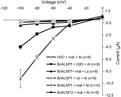
Figure 4.
Electrophysiological characterization of BnALMT1 and BnALMT2 protein in Xenopus oocytes. Oocytes were injected with cRNA of BnALMT1, BnALMT2, or water. After an overnight incubation at 20°C the oocytes were injected with 50 nL of 100 mm Na malate or Na citrate before being exposed to 100 μm AlCl3 or LaCl3. From the holding potential 0 mV, the test voltage was clamped from −100 to 0 mV in 20 mV increments (3 s pulses). After clamping the membrane potential at a test voltage, the voltage was maintained at the holding potential for 5 s before being clamped at the next test voltage. The current-voltage curves were constructed from the current values measured at the end of the 3 s voltage pulses.

