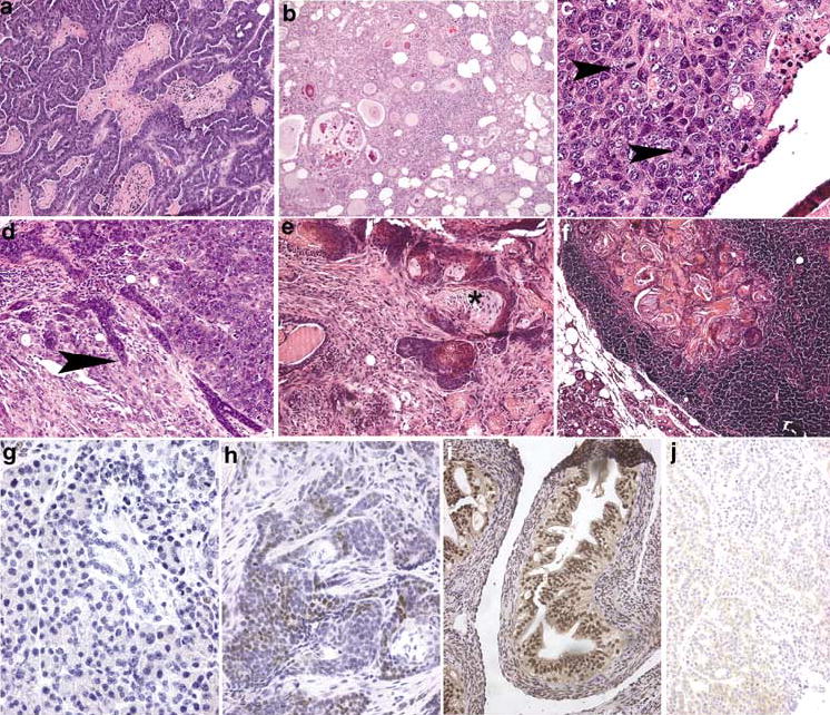
Figure 3.

Mammary neoplasia in NRL-PRL transgenic mice. (a) Papillary adenocarcinoma, with focal necrosis. (b) Glandular adenocarcinoma, in which neoplastic epithelial cells form glands that often contain eosinophilic secretions. (c) Solid carcinoma, showing nuclear pleomorphism and multiple, often abnormal, mitotic figures (arrowheads). (d) Solid carcinoma with focal invasion into adjacent stroma (arrowhead). (e) Adenosquamous neoplasm, containing clusters of cells with central squamous differentiation (asterisk). (f) Adenosquamous neoplastic cells present in a mammary lymph node. (g) ERα− mammary neoplasm. (h) ERα+ mammary neoplasm; note the brown stain present over many nuclei. (i) ERα+ control uterus. (j) ERα+ neoplasm treated with nonimmune serum as a negative control. Original magnification: (A, B, F), ×100; (D, E, I, J), ×200; (C, G, H), ×400
