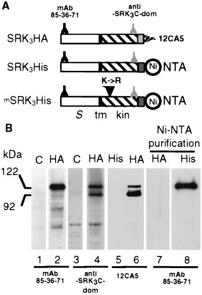
Figure 1.
Expression of recombinant SRK3 proteins in insect cells. (A) Schematic representation of the three recombinant SRK proteins, SRK3HA, SRK3His, and mSRK3His. The position of the different epitopes and tags are shown. The N and C termini of the recombinant proteins are to the left and right, respectively. The white rectangle represents the S-domain (S), the black vertical bar the membrane-spanning (tm) domain, the hatched rectangle indicates the cytoplasmic domain (kin) and the light and dark gray stippled rectangles indicate the HA epitope and hexahistidine tag, respectively. Epitopes recognized by different antibodies and the binding site for Ni-NTA are indicated. The substitution of Lys-553 with an arginine (K->R) in the mSRK3His construct is indicated by a vertical arrowhead. (B) Immunoblotting of SRK recombinant proteins. Proteins extracted from Sf21 cells infected by the parental baculovirus (C) (lanes 1 and 3) or from Sf21 cells infected with baculovirus driving the expression of SRK3HA (HA) (lanes 2, 4, 6, and 7) or SRK3His (His) (lanes 5 and 8) were separated by SDS/PAGE and electroblotted. In some cases, electrophoresis was preceded by a purification with Ni-NTA agarose beads as indicated.

