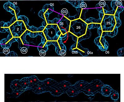
Figure 1.
Two sections of electron density at 1.0-Å resolution. (A) Segment of four glucoses, G25 to G2, showing one of the two bandflip sites in CA26 defined by G26-G1; these two glucoses are stabilized in anti orientation by the three-center hydrogen bond O(3)26—H⋅⋅⋅(O(5)1,O(6)1. The adjacent glucoses on both sides of the flip are oriented syn as usually found in amylose chains and hydrogen bonded O(3)n⋅⋅⋅O(2)n+1. Because the flip at G26-G1 involves not just a single glucose but the whole appended amylose chain, it was called “band-flip” (18). Labels of O(2) and O(3) hydroxyl groups are circled to emphasize the abrupt structural change at the band-flip site. (B) Disordered water molecules located in the channel-like cavity of the V-amylose helix. Because the distances between their positions (marked ∗) are shorter than the minimum hydrogen bonding distance of 2.5 Å (30), the occupations are around 0.5. Drawn with o (31).

