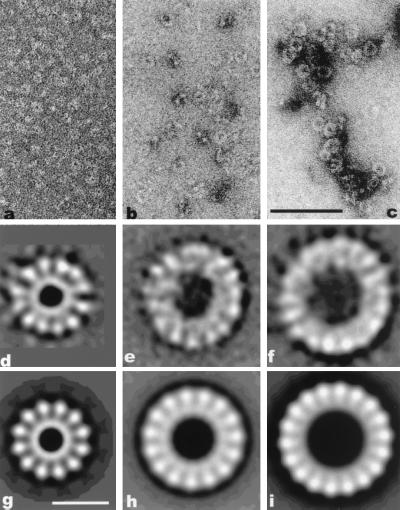
Figure 1.
(a–c) Electron micrographs of β protein forming small rings in the presence of MgCl2 (a), oligonucleotides (b), or ssM13 (c). (Scale bar = 1,000 Å.) (d–i) Image averages. (d) Average of 900 small rings showing 12 subunits. (e) Average showing ≈15 subunits from the 300 large rings with the strongest 15-fold rotational power. The rings were formed in the presence of a 30-mer oligonucleotide. (f) Average of 600 large rings with 18 subunits assembled after incubation with heat-denatured calf thymus DNA. (g) Twelve-fold symmetrized image from d. (h) Fifteen-fold symmetrized image from e. (i) Eighteen-fold symmetrized image from f. (Scale bar = 100 Å.)

