Figure 4.
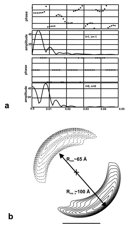
(a) Averaged layer lines from 34 filaments. (b) Cross section of the helical reconstruction generated from the layer lines in a. The reconstruction is shown by two slices, one at z = 0 Å (solid lines) and one at z = −62.5 Å (dotted lines), half of a helical turn away. The outer contour shown encloses a volume of 700,000 Å3 for one helical turn, and was chosen so as to yield a thickness (between Rmin and Rmax shown) of about 35 Å. This was the approximate thickness of the strands seen in regions (Fig. 2e) where the filaments unravel. The cross sections show that the helix consists of a single compact nucleoprotein strand, as shown in the model of Fig. 5b.

