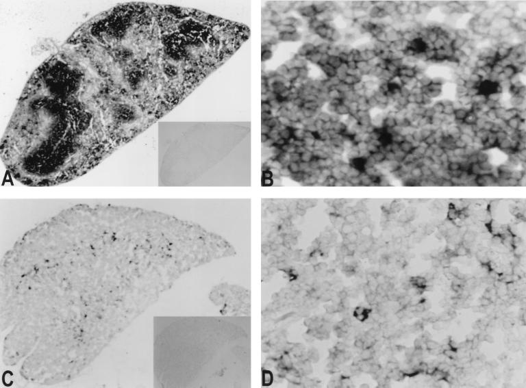
Figure 4.
In situ hybridization on mouse tissue sections. Mouse thin sections (5 μ) were hybridized with FAT10 antisense (A and B, spleen; C and D, thymus) and sense probes (insets in A and C). Expression of murine FAT10 mRNA was seen only with antisense probe. The heavily stained cells are scattered in the white pulp in the spleen (A and B). In mouse thymus, the positive cells were mainly in cortico-medullary junction (C). At higher resolution (D), most FAT10 mRNA was in cells with expanded cytoplasm.

